Radiography students' research displayed on informative posters
Wednesday, May 3, 2023
Photos by Wendy A. Miller, assistant dean of health sciences
Students in Allen R. Smith’s Radiographic Anatomy & Positioning IV class recently displayed research posters in The Madigan Library. The posters are a culmination of two months of work that builds on the knowledge students gained in class.
Working in randomly assigned groups of three or four, the 22 students could choose to research any topic related to fluoroscopy, which is the focus of the course. Fluoroscopy uses X-rays and contrast material (positive and negative) to provide real-time imaging to visualize a patient’s internal structures and functions. These real-time images are displayed on a video monitor computer screen.
Smith is clinical director of radiography.
Students in the Radiologic Anatomy & Positioning IV class shared information on a variety of topics related to fluoroscopy in the college’s Madigan Library.
Student posters explained the purpose of a procedure, what patients can expect and a radiographic technologist’s role, and addressed common questions.
Students researched:
- Hysterosalpingogram: An X-ray procedure that is used to visualize the uterus and fallopian tubes. It can be used to determine if either of the tubes is blocked and, sometimes, can be therapeutic and remove blockages.
“We want to bring more awareness to the importance of this procedure and how it helps women to be able to conceive,” the group wrote in its abstract.
Students: Erin R. Dean and Sofie A. Dean, both of Harrisburg; and Grace E. Pfleegor, of Williamsport.
- Radiation Protection: In fluoroscopy, X-ray is used as a guide while a procedure is taking place, so radiological technologists, radiologists and other medical personnel are in the room where the radiation exposure is being made. It is the radiographer’s duty to make sure everyone is properly equipped with protective gear.
“Radiation protection is a must to understand when dealing with fluoroscopy,” the group’s abstract states. “Fluoroscopy is real-time imaging, causing for longer exposures. We decided to choose this topic to prepare ourselves/others more for the levels of (radiation) exposure, the precautions we should be taking to reduce our dosage, and to have a better understanding of the equipment we use to help us reduce our exposure.”
Students: Alexis M. Auman, of Sunbury; Danielle L. Hoffman, of Slatington; Thomas L. Lewis III, of Moshannon; and Abigail M. Wisniewski, of Middletown.
- Barium Swallow: This procedure visualizes the gastrointestinal system (the mouth, esophagus and stomach). Barium, a contrast medium, is swallowed by a patient to show the functioning parts of the GI system.
“This is an important exam … because, often, it helps to figure out why a patient is having difficulty swallowing their food,” the students state. “Eating is a crucial part of survival, and it is critical for patients to understand why they may be struggling to consume their food.”
Students: Morgan Feeley, of Bridgewater, N.J.; Kaitlyn R. Hauck, of Hughesville; Jordan Palmer, of Honesdale; and Austin M. Rizzo, of Williamsport.
- Arthrograms: Arthrograms provide fine details of tiny structures inside joints, including the wrist, shoulder, hip and knee. X-ray contrast is injected to view the joint space. It can be used to find various issues, including ligament tears.
“Arthrograms are important to (show) what is happening inside the joints, without any incision, to explain joint pain,” the group’s abstract states.
Students: Kas F. Kondash, of Millheim; Alyssa A. Martz, of Middletown; Ashlee D.L. Robins, of Milton; and Megan M. Shaylor of Reading.
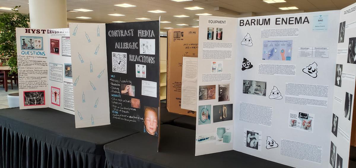
- Contrast Media Allergic Reactions: Allergic reactions can occur with either ionic or non-ionic contrast. These reactions can range from mild (nausea/vomiting, headache, itching or flushing) to moderate (skin rashes or hives, wheezing, high or low blood pressure, or shortness of breath), or severe (swelling or profound low blood pressure). These reactions are less likely to occur with non-ionic contrast compared to ionic contrast media.
“We chose to present the idea of allergies to contrast to inform other health care workers and the public of the safety risks it can cause. … Appropriate preventative measures taken before a contrast media exam help avert any allergic reactions that may occur,” the group’s abstract states. “Health care professionals should be educated accordingly when working with or close to contrast media.”
Students: Gabrielle N. Betts, of Montoursville; Kassidy A. Krammes, of Auburn; Kelsey J. Kronenwetter, of Williamsport; and Jessica L. Williams, of Williamsport.
- Barium Enema: Also known as lower GI series, a barium enema exam uses contract media to examine the large intestine. The exams are less common in fluoroscopy because CT scans provide better imaging and are less time-consuming. Common findings are Chron’s disease, diverticulitis, intussusception, ulcerative colitis and neoplasms.
“Barium enemas are known to be uncomfortable for all parties involved in the exam, but it is a very important exam to have done,” the students state. “Barium enemas are important because they are used to diagnostically find perforations as well as diagnose cancers and other harmful pathology.”
Students: Frederick Leland, of Mill Hall; Skylar J. Merrick and Mya L. Ott, both of Williamsport; and Elisabeth Shaffer, of Trout Run.
